IHC staining of cancer cases @Sapien

At Sapien Biosciences, we screen our biobanked FFPE samples using Ventana IHC systems for optimization and staining of antibodies. This allows to visualize the presence and localization of target proteins within tissue samples, supporting biomarker validation, disease characterization, and therapeutic target identification. Our digital pathology capabilities include whole-slide scanning at 40X magnification for advanced AI and ML applications.


For example, IHC staining to detect MUC1 expression. MUC1 is often overexpressed or aberrantly glycosylated in many types of cancers, including breast, ovarian, lung, and pancreatic cancers. Its presence and expression levels serve as a useful biomarker for tumor detection and characterization. MUC1 is also a promising target for cancer therapeutics, including vaccine development and monoclonal antibody therapies.

Cell Assays for Immuno-Oncology R&D

June is Cancer Immunotherapy Awareness Month. Immunotherapy is a type of cancer treatment that enhances the patient’s own immune system to recognize and kill cancer cells. Several immunotherapies have been approved for use in treating cancer, such as checkpoint inhibitors, CAR-T cell therapy, and therapeutic vaccines.
The development of immuno-oncology drugs requires understanding their activity such as their ability to increase T cell response and production of cytokine. In-vitro functional assays are essential to assess their efficacy, MOA, safety and toxicity, particularly for mono- or bi-specific antibodies designed to target and kill cancer cells by recruiting patient’s immune cells.
Such clinically relevant assays using human cells are used @Sapien, as 2D or 3D tumour spheroids, to test drugs alone or in combination with FDA-approved drugs such as Keytruda, in lung cancer or triple negative breast cancer cells. The left side graph below shows one such patient-derived model, where cells were co-cultured with activated PBMCs, to assess combinatorial killing effect of a client’s anti-cancer drug with the PD-1 inhibitor Keytruda.
The right side graph below shows the Dendritic cell-Mixed Lymphocyte Reaction (DC-MLR) assay. It is used to assess the immunomodulatory activity of drugs such as Cyclosporine A or Teriflunomide, or novel NCEs or NBEs, in PBMC-derived dendritic cells stimulated with superantigen SEB and co-cultured with PBMCs.

HRR testing including BRCA1/2 can guide therapy in Ovarian Cancer

Specific types of DNA damage, such as mismatches during replication, single- or double-strand DNA breaks, can result in the activation of specific signalling and repair cascades such as homologous recombination repair (HRR) in normal cells to repair the damage. However, deficiencies in these repair pathways result in the accumulation of DNA damage, genomic instability, and an increased risk of developing cancers, particularly breast and ovarian cancers. Hence, it is important to test for mutations in the repair genes including the well-known BRCA1, BRCA2, plus others such as RAD51C, RAD51D, PALB2, and ATM, to improve the selection of targeted therapies like PARP inhibitors namely Olaparib, Niraparib, Rucaparib, and Talazoparib.

To determine the potential for PARP inhibitors in Indian ovarian cancers, we have been sequencing our 1100+ ovarian cancer resection cases using a 43-gene HRR NGS panel. We have identified several cases with BRCA mutations, as well as other HRR pathogenic/likely-pathogenic mutations in ARID1A, ATR, ATM, CDK12, CHEK2, RAD51B, FANCC, and FANCE genes. In the DNA damage response signaling pathway shown below, these genes are marked with an asterisk to indicate mutations identified in our ovarian cancer samples. These oncogenic alterations may predict response to PARP inhibitors and guide the choice of therapy to improve ovarian cancer patient lives.

Histological Distribution of Sapien’s Ovarian Cancer Cases

Ovarian cancer is comprised of various tumour groups that are histologically distinct which is important to know for their treatment. The cancer may originate in one or both ovaries, as well as in the nearby fallopian tubes or peritoneum. Diverse types of ovarian cancers are categorized based on the originating cell’s name.

Epithelial ovarian carcinomas, known as cancerous epithelial tumours, are the most prevalent among ovarian cancers. The American Cancer Society states that approximately 85 to 90 percent of ovarian cancers originate from epithelial cells that encase the outer surface of the ovary. Sapien ovarian cancer data of 1445 cases also shows that 95% of cases constitute epithelial tumours while only 5% are non-epithelial tumours such as germ cell and stromal tumours. Among epithelial tumours, 55% are identified as serous, followed by endometroid, adeno, mucinous, and clear cell carcinomas. In contrast, 64% of non-epithelial tumours are germ cell tumors and 36% are stromal tumors.

BRCA1/2 testing can guide therapy in Ovarian Cancer

Homologous recombination repair (#HRR) pathway repairs double-strand DNA breaks, and its deficiency leads to Homologous recombination deficiency (#HRD), resulting in genomic instability and contributing to cancer.
HRD mutations, including #BRCA1 or #BRCA2 mutations are important actionable biomarkers in #Ovariancancer, that are sensitive to #PARP inhibitors that block the repair of #DNAdamage, such as #olaparib, #rucaparib, and #niraparib.

Patients with mutations in BRCA1/2 account for ~15% (range 7%–21%) of ovarian cancers. Several phase-III double blind, randomized trials have shown that patients with advanced ovarian cancer carrying BRCA1/2 mutations have better progression-free survival (PFS) with PARP inhibitors compared to placebo (SOLO-1 trial Olaparib, PAOLA-1 trial Olaparib + Bevacizumab, PRIMA trial Niraparib)

We sequenced 89 of our 1100+ Ovarian cancer resection cases using a 43-gene HRR #NGS panel, which identified several mutations in genes involved in HR pathway, such as TP53, BRCA1, ARID1A, BRCA2, PTEN, ATR and ATM. We identified pathogenic/likely pathogenic BRCA1/2 #missense and #truncated mutations (see figure below) that result in the loss of BRCA1/2 tumor suppressor function. These oncogenic alterations may predict response to PARP inhibitors and guide choice of therapy.

Today is World Ovarian Cancer Day

World Ovarian Cancer Day is observed on May 8th each year, to raise awareness about Ovarian Cancer, its symptoms, risk factors, and the importance of early detection and personalized treatment. Ovarian cancer is one of the most common gynecological cancers that ranks third after cervical and uterine cancer with high incidence in Asian women (9.2 per 100,000), with poor prognosis and high mortality rate.

In our biobank, we observe most ovarian cancer cases are detected at a young age with a median age of only 53 years, and in most cases cancer had already advanced and spread to both ovaries, bilateral cancer. If they had been detected earlier, they could have been cured as the cure rate for ovarian cancer is >80% if detected at early stage.

Epidemiology of ovarian cancer, June 2020
https://pdfs.semanticscholar.org/17c5/6233f2de1692f4b542db46bfec48e8090084.pdf

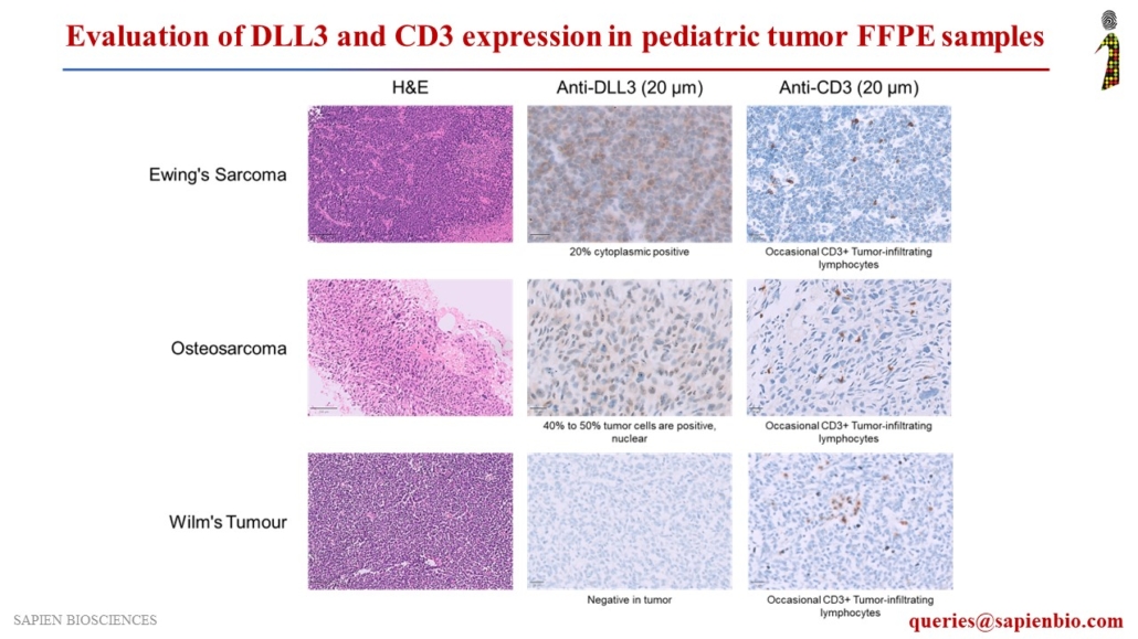
Assessment of DLL3 and CD3 Expression in Pediatric Tumor FFPE Samples for Potential DLL3-Targeted Immunotherapy

Delta-like ligand 3 (DLL3), a Notch inhibitory ligand, is a promising therapeutic target that is upregulated in Small Cell Lung Cancers (SCLC) and Neuroendocrine carcinomas but is not detectable in normal adult tissues. Various DLL3-specific therapies are under clinical development, including ADC rovalpituzumab tesirine (#Abbvie), bispecific TCE molecule AMG 757 (#Amgen), and CAR-T therapy AMG 119 (#Amgen), for the treatment of SCLC and neuroendocrine carcinomas.

We evaluated DLL3 expression in several tumor types, along with CD3, a marker for T-cell infiltration in tumors, to assess their potential for DLL3-targeted immunotherapeutic agents. DLL3 showed positive staining in neuroblastoma and pediatric tumors, such as Ewing’s sarcoma, osteosarcoma, rhabdoid tumors, Hodgkin’s lymphoma, rhabdomyosarcoma, acute myeloid leukemia (AML), and acute lymphoblastic leukemia (ALL).

The landscape of actionable genomic alterations in lung adenocarcinomas in India

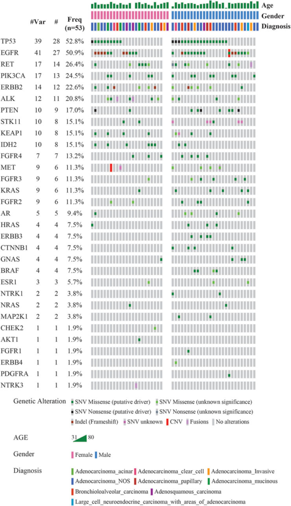
Sapien Biosciences is excited to share our recently published article, titled “The Landscape of Actionable Genomic Alterations in Lung Adenocarcinomas in India,” in Frontiers in Genetics, section Genetics of Common and Rare Diseases.

Our retrospective study, involving 53 Indian Lung Adenocarcinoma patients and utilizing a targeted NGS panel of 46 cancer-relevant oncogenes, revealed pathogenic or likely pathogenic variants in 94% of cases. Notable findings include a high prevalence of TP53 (52.8%) and EGFR (50.9%) mutations, gender-based differences in genetic alterations, and the detection of clinically actionable variants in 79% of cases. These results emphasize the crucial role of NGS in uncovering actionable molecular alterations, enhancing our understanding of NSCLC’s molecular landscape, and facilitating personalized treatment for Indian patients based on their unique genetic profiles.

We extend our gratitude to the entire research team, collaborators @Thermo Fisher Scientific, @q-annie-hasan, @Dr Aruna Priya Kamireddy, and Frontiers in Genetics for helping us make this significant contribution to Indian genomics research possible.

Please find the link to our publication below:

https://www.frontiersin.org/articles/10.3389/fgene.2023.1256756/full

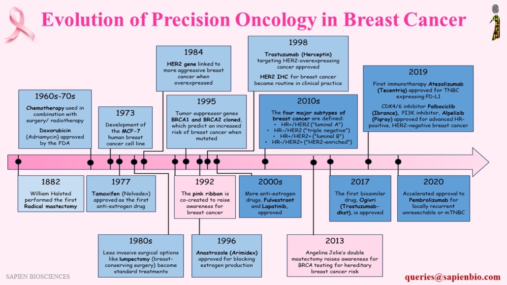
Timeline of Precision Medicine in Breast cancer

The timeline of breast cancer care and therapy is marked by significant milestones that have transformed patient outcomes and the way we approach this devastating disease as shown in the figure here. From the pioneering mastectomy procedures of the late 19th century to the development of targeted therapies specifically blocking the drivers of breast cancer such as ER, PR, HER2 personalized medicine has come a long way in recent years. The diagnosis and therapy guidance of breast cancer has evolved now to 4 major subtypes with personalized treatment strategies that are tailored to individual patient’s tumour characteristics and aggressiveness.

Breakthroughs in chemotherapy, hormonal therapy, most recently immunotherapy, along with advances in surgical techniques to preserve the breast tissue, and improvement in radiation therapy have all contributed to significant progress in breast cancer treatment. In the last 20 years, multiple treatment options have emerged for recurred and metastatic breast cancer. The effectiveness of such targeted treatments is higher and it has fewer side-effects, so it improves the quality of life. Simultaneously, awareness of risk factors including the role of BRCA1/BRCA2 genes have led to more women understanding hereditary risk and the need for regular screening.

In recent years, Next-Generation Sequencing (NGS) has been a pivotal player, enabling comprehensive genome analysis and uncovering rare genetic mutations that can be the targets of new drugs. These breakthroughs have ushered in an era of personalized medicine enhancing the overall quality of care as well as long-term survival in breast cancer.

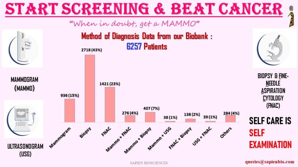
Diagnostic methods for breast cancer used in India

Breast cancer can be diagnosed through multiple tests, including a mammogram, self-examination, ultrasound, MRI and biopsy. If the doctor finds an area of concern on a screening test (a mammogram), or if the patient has symptoms that could mean breast cancer, then more tests need to be done in order to diagnose whether it is cancer or not. The most common symptom of breast cancer is a new lump or mass. Breast cancer is sometimes found after symptoms appear, but many women with breast cancer have no symptoms. This is why regular breast cancer screening is so important. Any change in breast should be checked by a physician.

Most widely used methods of diagnosis are as follows:
1) Mammograms : low-dose x-rays
2) Ultrasound (USG) : sound waves and their echoes to make computer pictures of the inside of the breast. It can show certain breast changes, like fluid-filled cysts, that can be harder to see on mammograms.
3) MRI : radio waves and strong magnets to make detailed pictures of the inside of the breast.
4) Biopsy including Core needle biopsy, FNAC, or Surgical biopsy: this is done when mammograms, or another imaging test, or a physical exam shows a breast change that may be cancer.
A biopsy is the only way to know for sure if it’s cancer.

References:
Breast Cancer Mode of Detection in a Population-Based Cohort- 2022-

https://pubmed.ncbi.nlm.nih.gov/36737116

Tru-Cut Biopsy versus Fine Needle Aspiration Cytology in Diagnosis of Early Breast Cancer-2023 https://ijmpr.in/article/Tru-Cut+Biopsy+versus+Fine+Needle+Aspiration+Cytology+in+Diagnosis+of+Early+Breast+Cancer


Fine-needle aspiration cytology versus core needle lymph node biopsy in axillary staging of breast cancer – 2022 https://ejrnm.springeropen.com/articles/10.1186/s43055-022-00895-w
Comparison of ultrasound and mammography for early diagnosis of breast cancer among Chinese women with suspected breast lesions: A prospective trial – 2022
https://www.ncbi.nlm.nih.gov/pmc/articles/PMC9663682/